Quantification of citrus-greening disease pathogens using competitive PCR
Description
[Objectives]
Huanglongbing (HLB, also known as citrus greening) is one of the most severe diseases affecting citrus production in tropical and sub-tropical regions of the world, and has recently spread to citrus orchards in southern areas of Japan. Diagnosis of this disease is usually conducted through symptom observation and PCR detection, but these methods do not allow the quantification of pathogen numbers.
The quantification of pathogens is crucial for analyzing the process of disease development. Based on recent research results, we have developed a quantification system for Huanglongbing pathogens, utilizing a competitive PCR method. This system consists of conventional PCR equipment and competitor DNA, and is less expensive than equipment used in real-time PCR systems, which currently is the most popular method for quantification of PCR-detectable materials. Thus, this system will be more easily incorporated into agricultural research activities in developing countries.
[Results]
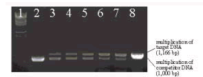
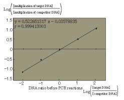
The conventional PCR method multiplies specific target DNA through chain reactions. The concept of the competitive PCR method is to create competition between target DNA multiplication and competitor DNA multiplication. The ratio of multiplied DNA from target DNA and competitor DNA is expected to correlate with the original ratio of each type of DNA before multiplication. We designed competitor DNA that has PCR-primer recognition sequences identical to target DNA. Using this competitive DNA, the competitive PCR reaction showed reasonable results (Fig. 1). After plotting data on a logarithmic graph, the multiplication ratios of each DNA formed nearly perfect straight lines (Fig. 2). Based on these results, the quantity of target DNA, pathogenoriginated DNA, as well as the estimated quantity of disease pathogens can be calculated.
There are several substances known to interfere with PCR DNA multiplication. Quantification systems using competitive PCR are thought to be less affected by such inhibitors. Starch is known to have accumulated in Huanglongbing infected leaves, and have inhibitory effects against PCR multiplication. We tested the quantification system with soluble starch in order to examine the reliability of measured values in the presence of such inhibitory substances. Favorable results were obtained from our quantification system as expected based on the concepts of the competitive PCR method.
Infected citrus leaves collected in southern Vietnam were applied in this quantification system, and significant differences in pathogen quantity were observed in leaves between two selected areas (Table 1). Fewer amounts of pathogen-related DNA were detected from the tissue of citrus cultivars that were believed to havehigher tolerance against Huanglongbing than that of susceptible cultivars. This suggests that the suppression of pathogen multiplication in tolerant citrus cultivars may enhance its tolerance.
We plan to utilize this quantification system in analyzing Huanglongbing disease prevalence, both in Japan and Vietnam, in a new international project to be initiated in April 2004 entitled, “Development of new technologies for control of Citrus Huanglongbing (HLB) in Southeast Asia.”
Figure, table
-
Fig. 1. An example of competitive PCR.
Lane 1: DNA size markers (λ- Eco T141 digest).
Lane 2: Multiplication of competitor DNA. Lane 3-7: 1pM target DNA and competitor
DNA, concentrations of each lane are 100pM, 10pM, 1pM, 100nM, and 10nM, respectively.
Lane 8: Multiplication of target DNA. -
Fig. 2. Competitive reactions between target DNA and competitor DNA. -
Table 1. Quantity of pathogens in infected citrus leaves.
- Affiliation
-
Japan International Research Center for Agricultural Sciences Okinawa Subtropical Station
- Classification
-
Technical A
- Term of research
-
FY2003 (FY2002-2005)
- Responsible researcher
-
KAWABE Kunimasa ( Okinawa Subtropical Station )
- ほか
- Publication, etc.
-
Kawabe, K., Truc, N. T. N., Nhan, N. T. and Onuki, M. (2004): Pathogen quantification of citrus tissue naturally infected by Huanglongbing in southern Vietnam. Jpn. J. Phytopathol., 70(3), 294
- Japanese PDF
-
2003_32_A3_ja.pdf2.71 MB
- English PDF
-
2003_32_A4_en.pdf51.64 KB


